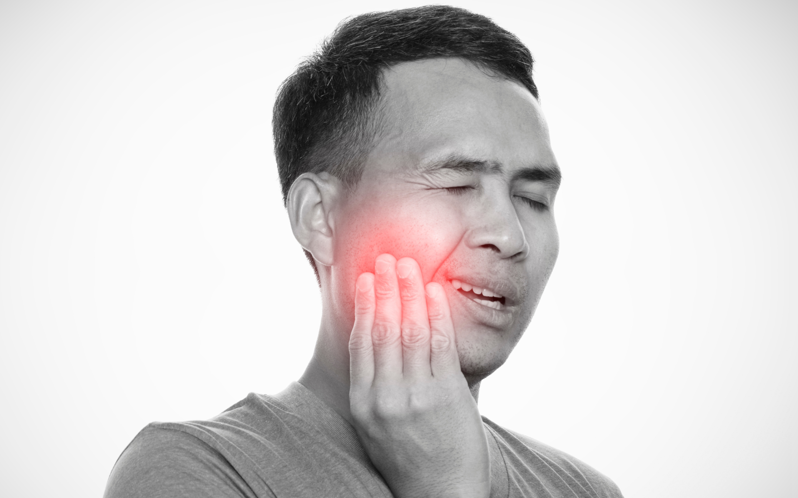
Your Jaw Pain Has a Story. Here’s How to Read It.
TMJ pain is one of the most commonly misunderstood and undertreated conditions we see at Voltex Physical Therapy in Austin. Patients come to us after months — sometimes years — of chasing symptoms: a night guard from the dentist, anti-inflammatories from their primary care doctor, maybe a referral to a specialist who mentioned Botox or surgery. The jaw got all the attention. The rest of the system got ignored.
The problem is that TMJ pain is rarely just a jaw problem. It involves a complex interplay of muscles, joints, nerves, and cervical spine mechanics that most treatment approaches address incompletely — or don’t address at all. To understand why you hurt and what will actually help, you need to understand the full picture: what type of TMJ disorder you have, which muscles are driving your symptoms, and why your neck is almost certainly part of the story.
That’s what this article is for.
What Is the TMJ — and What Can Go Wrong?
The temporomandibular joint is the hinge and gliding joint that connects your lower jaw (mandible) to your skull (temporal bone) on both sides of your face. It is one of the most complex joints in the body — capable of rotation, translation, lateral deviation, protrusion, and retraction, often simultaneously. It is involved in chewing, speaking, swallowing, yawning, and breathing. You use it hundreds of times per day without thinking about it.
When it works well, you never notice it. When it doesn’t, you can’t ignore it.
Temporomandibular disorders (TMD) is the umbrella term for a group of conditions affecting the TMJ, the muscles of mastication, and associated structures. According to the Diagnostic Criteria for Temporomandibular Disorders (DC/TMD) — the current international gold standard for classification — TMD is divided into two primary categories: pain-related disorders and intra-articular (structural) disorders. Many patients have both simultaneously.
Peck CC, et al. (2021). Temporomandibular disorders: Current Concepts and Controversies in Diagnosis and Management. PMC. https://pmc.ncbi.nlm.nih.gov/articles/PMC8000442/
Why Does Sleep Bruxism Happen? The Neuroscience Behind the Grind
Bruxism is not simply a bad habit you can decide to stop. Its etiology is multifactorial — driven by a complex interaction of neurological, psychological, physiological, and lifestyle factors. Understanding why your brain is activating your jaw muscles during sleep is the key to understanding why a passive dental appliance, on its own, rarely resolves it.
The Role of the Central Nervous System
Sleep bruxism is now understood to be primarily a centrally mediated behavior — meaning it originates in the brain, not in the jaw itself. During sleep, the jaw muscles receive rhythmic activation signals from the central nervous system, likely related to normal sleep arousal mechanisms. In people with bruxism, these signals are amplified or dysregulated, producing the forceful, prolonged contractions that cause morning pain.
Research has consistently linked sleep bruxism to dopaminergic pathways in the brain — the same neurotransmitter system involved in stress response, movement regulation, and reward. This is part of why certain medications (particularly SSRIs, stimulants, and some antipsychotics) can worsen bruxism, and why stress is such a reliable trigger.
Stress, Anxiety, and the Austin Factor
Psychological stress is one of the most well-documented triggers for both sleep and awake bruxism. Elevated life stress and anxiety increase nocturnal jaw muscle EMG activity — meaning a stressful week at work, a demanding project, or a period of anxiety directly translates into harder, more frequent clenching during sleep. For Austin’s high-achieving, high-stress professional population, this connection is clinically relevant and frequently underappreciated.
The masseter muscle, in particular, is one of the primary muscles the body recruits in response to psychosocial stress. When the nervous system is chronically activated — whether from work pressure, device overuse, poor sleep, or ongoing pain — the jaw becomes a preferential tension storage site. Treating bruxism without addressing the nervous system load is treating the output, not the input.
Sleep Disorders: The Connection Most Patients Miss
An important and underrecognized driver of sleep bruxism is obstructive sleep apnea (OSA). Research has documented a significant association between sleep bruxism and OSA — with the prevailing theory being that bruxism episodes may serve as a protective mechanism to re-open the airway during partial obstruction. Patients who present with severe morning jaw pain, combined with daytime fatigue, snoring, or reported apneic episodes, warrant evaluation for OSA before bruxism treatment is finalized.
If your bruxism hasn’t responded to conservative care, ask your provider whether a sleep study is appropriate. Treating the bruxism without treating the underlying sleep disorder will produce incomplete and frustrating results.
Uchima Koecklin et al. (2024). The neural substrates of bruxism: current knowledge and clinical implications. PMC. https://pmc.ncbi.nlm.nih.gov/articles/PMC11473305/
Kuang B, et al. (2022). Associations between sleep bruxism and other sleep-related disorders in adults: a systematic review. Sleep Med, 89:31–47.
The Types of TMJ Pain: What’s Actually Happening in Your Jaw
Not all jaw pain is the same — and the type of pain you have determines which structures are involved, which treatments are most appropriate, and what your recovery should look like. The DC/TMD identifies twelve of the most common TMD diagnoses. Here are the ones most relevant to what we treat at Voltex PT in Austin:
1. Myofascial Pain (with and without Referral)
This is the most common type of TMJ-related pain — and the one most directly addressed by physical therapy. Myofascial pain originates from trigger points in the muscles of mastication: the masseter, temporalis, medial pterygoid, and lateral pterygoid. These hyperirritable spots in the muscle tissue produce local pain and — critically — referred pain that can travel to distant sites.
Masseter trigger points, for example, commonly refer pain to the teeth, ear, and cheek. Temporalis trigger points refer to the temple, the forehead, and behind the eye. Patients are frequently told they have ear infections, migraines, sinus problems, or dental issues when the actual source is a trigger point in a jaw muscle. Myofascial pain with referral is categorized separately from local myalgia precisely because the pain experience extends well beyond the muscle itself.
Clinical note: if you’ve been told your teeth look fine, your ear drum is normal, and your sinus X-ray is clear — but you still have face, ear, or head pain — myofascial trigger points in the jaw and neck muscles should be high on the differential.
2. Arthralgia — True Joint Pain
Arthralgia refers to pain that originates directly from the temporomandibular joint itself — specifically from the joint capsule, synovial membrane, and retrodiscal tissue (the highly vascularized and innervated tissue behind the articular disc). Unlike myofascial pain, which can travel widely, arthralgia is felt directly in the joint and is typically reproduced by direct palpation of the joint or by jaw movement.
Arthralgia frequently co-exists with disc displacement and inflammatory joint changes. Patients describe a deep ache in front of the ear, often worse in the morning or after prolonged jaw use. While arthralgia involves true joint pathology, physical therapy can significantly reduce pain by decreasing joint compression, improving disc mechanics, and reducing the muscular forces that load the joint.
3. Disc Displacement — The Source of Clicking and Locking
The articular disc is a fibrocartilage cushion that sits between the condyle and the temporal bone, normally moving forward and backward as the jaw opens and closes. In disc displacement with reduction, the disc slips forward out of position but snaps back (reduces) during mouth opening — producing the characteristic clicking or popping sound that many TMJ patients know well. In disc displacement without reduction, the disc remains displaced and blocks full jaw opening, causing the locking and limited mouth opening that can be acutely alarming.
Disc displacement affects an estimated 28–35% of the general population in some form, though it does not always cause pain. When it does produce pain, it is typically through the compression of the retrodiscal tissue and altered loading of the joint surfaces. Physical therapy can improve disc mechanics, reduce compressive forces, and in many cases restore more normal opening without intervention.
Emedicine/Medscape. Temporomandibular Joint (TMJ) Syndrome: Pathophysiology, Epidemiology. https://emedicine.medscape.com/article/809598-overview
4. Degenerative Joint Disease (TMJ Osteoarthritis)
TMJ osteoarthritis is a slowly progressive degenerative condition characterized by cartilage degradation, subchondral bone remodeling, and eventual bony changes visible on imaging — including condylar erosion, flattening, and osteophyte formation. It is more prevalent in females and increases with age, though it can present in younger patients with a history of trauma or significant bruxism.
Importantly, degenerative joint changes on imaging do not always correlate with pain. Many patients with significant structural changes have minimal symptoms; others with mild imaging findings have severe pain. This is why treatment should be guided by symptoms and function, not imaging alone — and why physical therapy targeting muscular load, joint mechanics, and movement patterns can provide meaningful relief even in the presence of degenerative change.
5. Headache Attributed to TMD
The DC/TMD formally recognizes headache as a distinct TMD diagnosis — specifically temporal headache that is aggravated by jaw function and reproduced by palpation of the masticatory muscles. This is distinct from tension-type headache or migraine, though TMD-related headache is frequently mislabeled as one of these. The masseter and temporalis muscles refer pain directly into the temples, forehead, and behind the eyes — creating headache patterns that resolve when the underlying muscular drivers are treated.
Physiopedia. Temporomandibular Disorders. https://www.physio-pedia.com/Temporomandibular_Disorders
The Muscles of the TMJ: Who Does What
To understand TMJ pain, you have to understand the muscular system that controls the jaw. There are four primary muscles of mastication — all innervated by branches of the trigeminal nerve (CN V) — and several accessory muscles that assist with jaw opening and stabilization. Each has a distinct role, a distinct pain referral pattern, and a distinct clinical significance.
Primary Muscles of Mastication
Masseter
Primary motion: Jaw elevation (closing). The most powerful muscle of mastication — responsible for the crushing force of chewing.
Clinical significance: The most commonly overloaded muscle in TMD. Hypertrophy from bruxism is visible as jaw widening. Trigger points refer to the cheek, teeth, ear, and eye. Nearly always involved in symptomatic TMD. Responds extremely well to dry needling.
Temporalis
Primary motion: Jaw elevation and retraction. The anterior fibers close the jaw; the posterior fibers retract the mandible.
Clinical significance: Trigger points in the temporalis are a primary driver of TMJ-related headache — referring pain to the temples, forehead, and behind the eyes. Palpable along the temporal fossa. Frequently involved alongside the masseter in bruxism and clenching patterns.
Medial Pterygoid
Primary motion: Jaw elevation and lateral deviation (contralateral). Works with the masseter to close the jaw and stabilizes the TMJ medially.
Clinical significance: Deep-seated muscle, difficult to palpate externally. Trigger points refer pain inside the mouth, throat, and behind the ear — often confused with ear infection or throat pathology. Frequently co-involved with the masseter in clenching. Accessible via intraoral techniques.
Lateral Pterygoid
Primary motion: Jaw depression (opening), protrusion, and lateral deviation. The only jaw muscle that actively opens the mouth and pulls the disc and condyle forward during opening.
Clinical significance: The most commonly implicated muscle in disc displacement. When the superior head of the lateral pterygoid becomes hyperactive or shortened, it pulls the disc anteriorly — which is the primary mechanism of disc displacement with reduction (clicking). Trigger points refer pain directly into the TMJ and behind the eye.
Accessory Muscles of Mastication
Several additional muscles assist with jaw opening and stabilization, and are frequently involved in TMD — particularly when cervical spine dysfunction is also present:
Digastric (Anterior and Posterior Bellies)
Primary motion: Jaw depression (opening) and hyoid stabilization. Works with the lateral pterygoid to open the mouth, particularly against resistance.
Clinical significance: The posterior belly is innervated by the facial nerve (CN VII) and is directly connected to the stylohyoid — bridging the jaw and the upper cervical region. Trigger points refer pain to the lower teeth and under the chin. Frequently involved in patients with forward head posture.
Suprahyoid Group (Mylohyoid, Geniohyoid, Stylohyoid)
Primary motion: Jaw depression and hyoid elevation. Stabilizes the floor of the mouth and coordinates swallowing with jaw movement.
Clinical significance: These muscles share innervation with upper cervical spinal nerves (C1–C3), creating a direct neuromuscular link between the jaw and the neck. Overactivation in TMD can increase compressive load on the cervical spine.
Infrahyoid Group (Sternohyoid, Sternothyroid, Thyrohyoid, Omohyoid)
Primary motion: Hyoid depression and cervical stabilization. Anchors the hyoid bone inferiorly, allowing the suprahyoids to generate jaw-opening force.
Clinical significance: Dysfunction in the infrahyoids alters the biomechanics of jaw opening and can contribute to cervical compression. The omohyoid descends all the way to the scapula — connecting jaw mechanics to shoulder girdle function.
National Academies of Sciences. (2020). Temporomandibular Disorders: Masticatory System Anatomy and Function. https://www.ncbi.nlm.nih.gov/books/NBK557988/
StatPearls. (2023). Anatomy, Head and Neck, Mastication Muscles. https://www.ncbi.nlm.nih.gov/books/NBK541027/
StatPearls. (2025). Anatomy, Head and Neck, Temporomandibular Joint. https://www.ncbi.nlm.nih.gov/books/NBK538486/
The jaw muscle map tells us something important: jaw mechanics and neck mechanics are not separate systems. They share innervation, share bony attachments through the hyoid, and are regulated by the same upper cervical neural circuitry. You cannot fully rehabilitate the jaw without addressing the neck.
The Neck and the Jaw: An Inseparable Relationship
One of the most underappreciated aspects of TMJ dysfunction is how frequently the cervical spine is involved — and how profoundly that involvement shapes the patient’s symptoms. At Voltex PT in Austin, treating the neck in TMD patients isn’t an add-on. It’s foundational.
The Numbers: How Common Is Cervical Involvement in TMD?
The research is striking. A study examining neck musculature pain patterns found that approximately 59% of TMD patients reported varying degrees of pain in the neck muscles — and that the severity of neck muscle involvement correlated directly with the severity of TMD. Another study found that cervical spine segmental limitations — particularly at the C0–C3 levels — and tender points in the sternocleidomastoid and upper trapezius were significantly more common in TMD patients than in controls.
In patients with existing cervical spine pathology, the numbers climb even higher: in a cohort of patients with cervical disc herniation, 88.5% of those with no or mild neck disability met criteria for TMD — and in those with moderate to severe neck disability, the rate was 100%.
Comparative Evaluation and Correlation of Pain Pattern in Neck Musculature in TMD Patients. PMC. https://www.ncbi.nlm.nih.gov/pmc/articles/PMC9642967/
Altunalan T, et al. (2025). Temporomandibular disorders and neck disability in individuals with cervical disc herniation. European Journal of Oral Sciences. https://pubmed.ncbi.nlm.nih.gov/41157797/
Why the Neck and Jaw Are Neurologically Linked: The Trigeminocervical Nucleus
The anatomical explanation for the jaw-neck connection lies in the trigeminal cervical nucleus — a convergence zone in the brainstem where pain signals from the jaw and face (carried by the trigeminal nerve, CN V) and pain signals from the upper cervical spine (carried by C1–C3 spinal nerves) are processed together. Because these inputs converge on the same neurons, the brain can misidentify where the pain is actually coming from.
This is why cervical spine dysfunction — a restricted C1–C2 joint, a trigger point in the suboccipital muscles, or a hypertonic sternocleidomastoid — can produce genuine facial pain, ear pain, and jaw aching that feels exactly like TMJ pain. It also explains the reverse: why an inflamed TMJ or overloaded masseter can refer pain into the neck, the occiput, and the shoulders.
The two regions are not just biomechanically connected — they are neurologically wired together. Treating one without the other is treating half the problem.
Neural Basis of Etiopathogenesis and Treatment of Cervicogenic Orofacial Pain. PMC. 2022. https://pmc.ncbi.nlm.nih.gov/articles/PMC9611820/
Effectiveness of Physiotherapy in TMJ Dysfunction and the Relationship with Cervical Spine. PMC. 2022. https://pmc.ncbi.nlm.nih.gov/articles/PMC9687864/
The Cervical Muscles Most Involved in TMD
Beyond the primary muscles of mastication, the following cervical muscles are frequently overloaded, hypertonic, or harboring trigger points in patients with TMD:
Sternocleidomastoid (SCM)
Primary motion: Head rotation, lateral flexion, and cervical flexion against gravity. Innervated by the spinal accessory nerve (CN XI) and C2–C3.
TMD relevance: SCM trigger points produce one of the most clinically significant pain referral patterns in the body — referring to the jaw, ear, eye, forehead, and top of the head. SCM hypertonicity is among the most common cervical findings in TMD patients. It is also directly affected by forward head posture, which loads both the SCM and the masticatory system simultaneously.
Upper Trapezius
Primary motion: Scapular elevation and cervical lateral flexion. Innervated by CN XI and C3–C4.
TMD relevance: Upper trapezius trigger points refer pain to the temple, the lateral neck, and the TMJ region — closely mimicking primary TMJ pain. Frequently chronically overloaded in desk workers and those with forward head posture, making it a near-universal finding in Austin’s tech and professional population.
Suboccipital Group (Rectus Capitis Posterior Major/Minor, Obliquus Capitis)
Primary motion: Craniovertebral extension, rotation, and fine postural control of the head on C1.
TMD relevance: Suboccipital restriction at C0–C1 and C1–C2 limits the normal coupling between head movement and jaw mechanics. The suboccipitals are also a primary source of tension-type headache and cervicogenic dizziness — both common TMD comorbidities. Direct connections between the suboccipital musculature and the dura mater explain why C1–C2 dysfunction can produce widespread craniofacial symptoms.
Splenius Capitis and Semispinalis Capitis
Primary motion: Cervical and capital extension, bilateral head extension, and ipsilateral rotation.
TMD relevance: Trigger points in the splenius capitis refer pain to the vertex of the skull and behind the eye. These muscles are routinely found to be hypertonic in TMD patients assessed under the DC/TMD protocol’s cervical examination criteria.
Caring Medical. TMJ: The Other Symptoms — Neck Pain, Muscle Spasms, Myofascial Pain. Trigeminocervical nucleus convergence. https://caringmedical.com
Craniocervical Misalignment Masquerading as Facial Pain. CRANIO Journal. 2020. https://www.tandfonline.com/doi/full/10.1080/08869634.2020.1823795
Forward head posture is the postural pattern we see most often in Austin’s desk-working, device-using population — and it is one of the most significant contributors to both TMD and cervical spine pain. For every inch the head translates forward from neutral, the effective weight on the cervical spine approximately doubles. This compressive load is transmitted directly into the suboccipital musculature, the upper traps, the SCM, and — through the hyoid and suprahyoid chain — into the jaw itself.
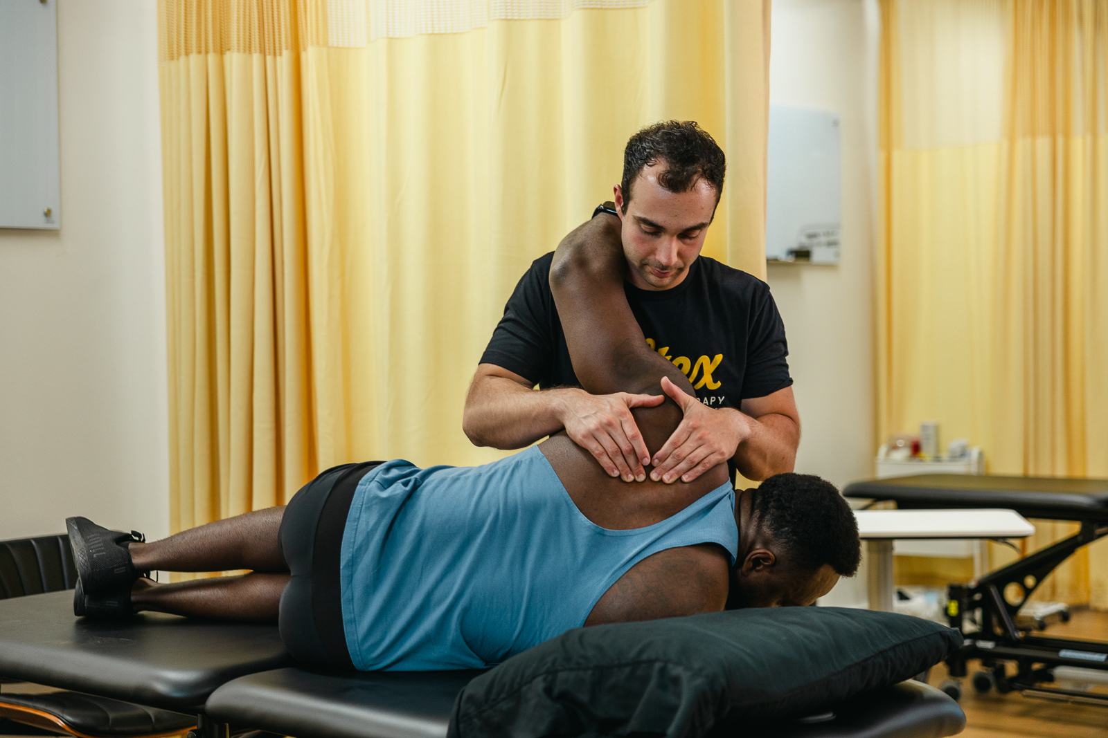
How Voltex PT Treats TMJ Dysfunction in Austin
At Voltex Physical Therapy on North Lamar in Austin, our approach to TMJ dysfunction is built on one foundational principle: treat the whole system, not just the joint that hurts. That means assessing and treating the jaw, the cervical spine, the hyoid complex, the masticatory muscles, the postural chain, and the movement patterns that are loading the system — all in the same treatment episode, from the same clinician, with your full undivided attention.
Here is how that translates into actual treatment:
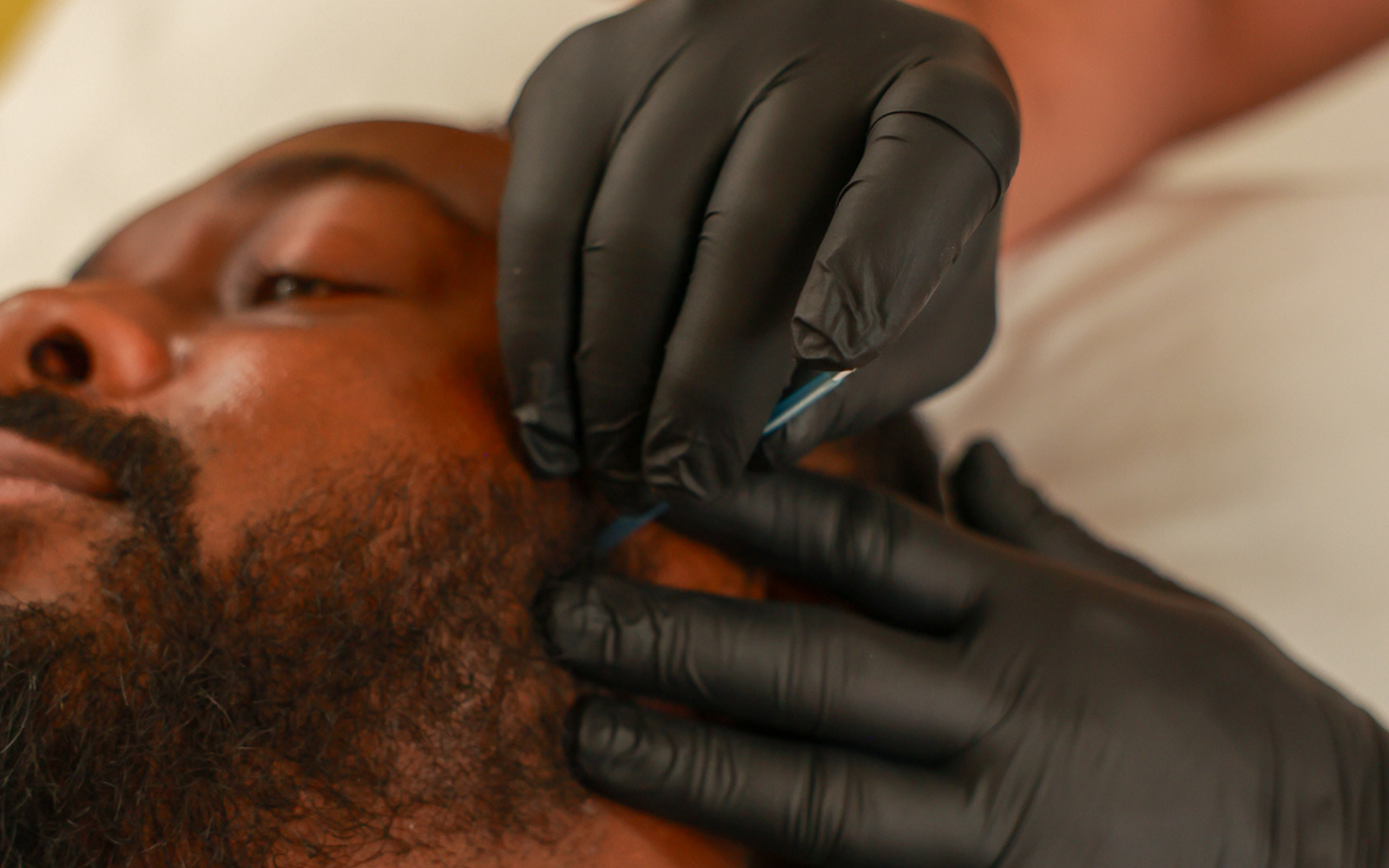
Dry Needling: Directly Targeting the Muscles Driving Your Pain
Trigger point dry needling is one of the most direct and effective tools we have for TMJ dysfunction — and the research consistently demonstrates why. By inserting a thin monofilament needle directly into a trigger point in the masseter, temporalis, pterygoids, SCM, suboccipitals, or upper trapezius, we elicit a local twitch response that immediately begins to reset the neuromuscular system: reducing muscle tone, improving local blood flow, decreasing sensitization of the muscle’s motor endplate, and allowing the muscle to return to normal resting length.
For patients with myofascial TMD, this can produce dramatic and rapid pain reduction — sometimes within the first session. For patients with more complex presentations involving disc dysfunction or articular pain, dry needling of the surrounding musculature reduces the compressive load on the joint and creates the mechanical environment in which other manual techniques can work more effectively.
Key muscles we needle in TMJ treatment at Voltex PT include:
Masseter — the primary jaw closer and the most commonly overloaded TMD muscle
Temporalis — for temple headaches, jaw tightness, and referral into the forehead
Medial pterygoid — for deep jaw pain, throat referral, and clenching-dominant presentations
Lateral pterygoid — for clicking, disc displacement mechanics, and anterior jaw deviation
Digastric — for under-chin tension and jaw-opening restriction
Sternocleidomastoid — for ear pain, facial referral, and cervicogenic headache
Suboccipital group — for C0–C2 restriction, occipital headache, and dizziness
Upper trapezius — for temporal referral and shoulder girdle involvement
Splenius capitis and semispinalis — for deep cervical tension and headache referral to the vertex
Dry needling does not replace manual therapy or exercise — it creates the window in which those treatments can work more effectively by reducing the baseline muscular hypertonicity that was limiting progress.
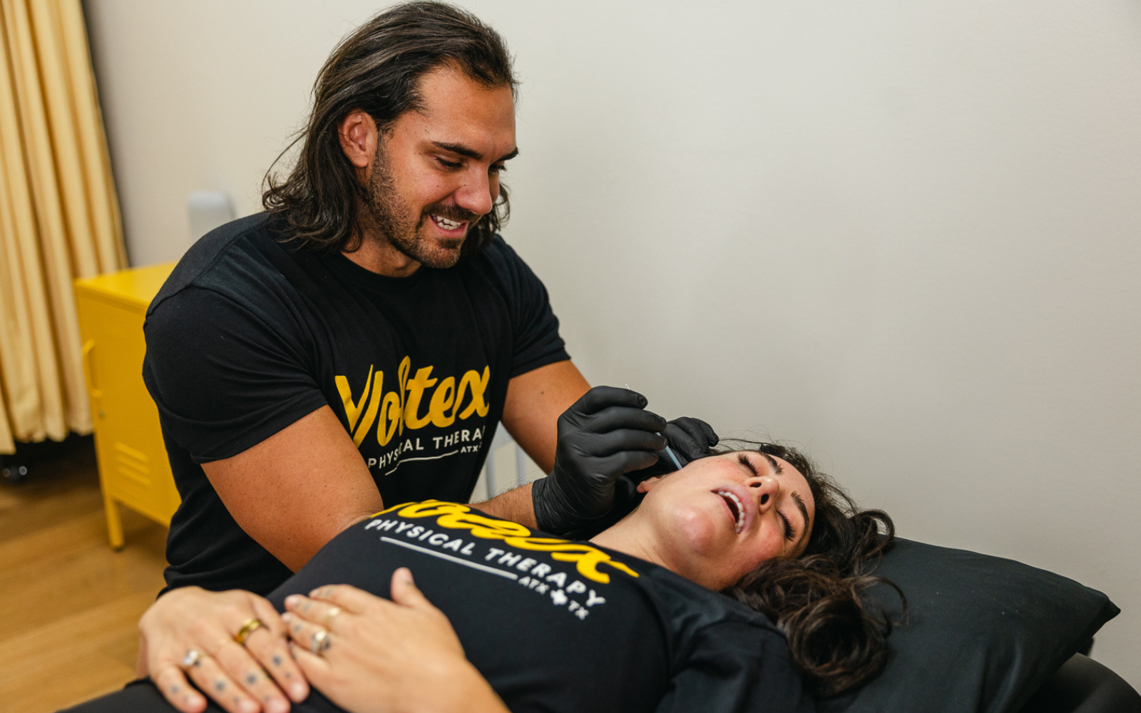
Manual Therapy: Restoring Joint Mechanics and Tissue Mobility
Manual therapy at Voltex PT for TMJ patients includes both hands-on work to the jaw itself and — critically — treatment of the cervical spine. These are not separate treatment components. They are applied as an integrated approach because the evidence, and our clinical experience with hundreds of Austin TMD patients, consistently shows that treating both produces far better outcomes than treating either in isolation.
Jaw-specific manual therapy techniques we use include:
Distraction and lateral glide mobilization of the TMJ to restore disc mechanics and reduce joint compression
Intraoral soft tissue release of the medial pterygoid and lateral pterygoid — accessible only from inside the mouth
Myofascial release to the masseter, temporalis, and surrounding facial fascia
Hyoid mobilization and suprahyoid soft tissue work to address the jaw-neck muscular bridge
Cervical manual therapy techniques we apply include:
C0–C1 and C1–C2 joint mobilization (Maitland grade III–IV) to restore craniovertebral mobility and reduce trigeminocervical sensitization
C2–C4 segmental mobilization for the mid-upper cervical levels most commonly restricted in TMD patients
Posterior cervical soft tissue release including suboccipital release and cervical paraspinal work
SCM and scalene soft tissue techniques to reduce cervical compressive loading
Thoracic extension mobilization, because upper thoracic restriction is a common driver of compensatory cervical tension
Vieira LS, et al. (2023). The Efficacy of Manual Therapy Approaches on Pain, Maximum Mouth Opening and Disability in Temporomandibular Disorders: A Systematic Review of 20 RCTs. Life. 13(2):292. https://doi.org/10.3390/life13020292
Upper Cervical Mobilization/Manipulation on TMJ Pain: Systematic Review and Meta-Analysis. PMC. 2023. https://pmc.ncbi.nlm.nih.gov/articles/PMC10036235/
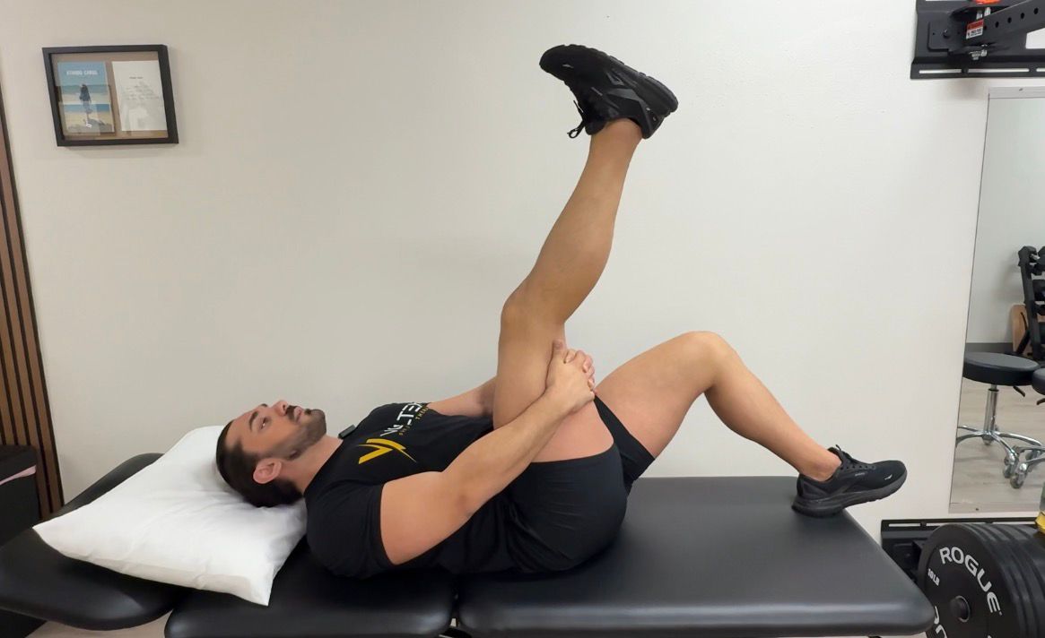
Postural Correction and Therapeutic Exercise
Pain relief is step one. Staying pain-free requires addressing the underlying patterns that loaded the system in the first place. For most TMD patients in Austin, that means correcting the forward head posture and cervical compression that has been feeding into their jaw pain — often for years.
Our exercise programming for TMJ patients typically includes:
Deep cervical flexor activation (longus colli, longus capitis) to restore cervical stability and reduce forward head posture
Scapular retraction and lower trapezius strengthening to open the thoracic spine and reduce upper trap dominance
Jaw proprioception and re-patterning exercises — controlled jaw opening with symmetry training to normalize condylar path
Diaphragmatic breathing retraining — mouth breathers and those with chronic respiratory patterns create paradoxical loading on the jaw and cervical spine
Postural load management: ergonomic guidance for desk setup, screen height, phone use, and sleep positioning — all of which directly affect the muscular load on the cervicalmandibular system
Patient Education: The Missing Piece
At Voltex PT, we consider patient education a treatment modality — not an afterthought. TMD patients need to understand what is driving their pain, what behaviors are perpetuating it, and what they can do between sessions to accelerate recovery. That includes:
Understanding the jaw-neck relationship so they stop treating their ‘jaw problem’ in isolation
Identifying and modifying parafunctional habits: clenching, gum chewing, teeth resting contact, jaw tensing during exercise or stress
Sleep hygiene as it relates to bruxism — because most nocturnal clenching is not fixed by a night guard alone
Stress load management, because the masseter is one of the primary muscles that responds to psychosocial stress — and Austin is a high-stress city
The Voltex PT difference: every session is one-on-one with a doctoral-level PT for the full session. No aides, no group exercises, no being handed a printout and sent to a gym corner. You get our undivided clinical attention — because jaw pain is complex, and it deserves complex thinking applied to it consistently.
Frequently Asked Questions: TMJ Pain and Physical Therapy in Austin
Is my jaw clicking dangerous?
Clicking or popping on its own, without pain or limitation in mouth opening, is usually not a medical emergency. It most commonly indicates disc displacement with reduction — the disc is slipping out of position and reducing back during jaw movement. Left unaddressed, clicking can progress to locking or pain in some patients, which is why it’s worth getting evaluated before symptoms escalate. A physical therapy evaluation can tell you which category you’re in and what, if anything, needs to be done.
Can physical therapy really help with TMJ — or do I need surgery?
For the vast majority of TMD presentations, physical therapy is the most appropriate and most effective first-line treatment — and surgery is rarely indicated. Evidence consistently supports conservative management including manual therapy, dry needling, and therapeutic exercise as effective interventions for pain reduction, improved mouth opening, and long-term function. Surgery is typically reserved for cases with severe structural pathology that has failed comprehensive conservative care.
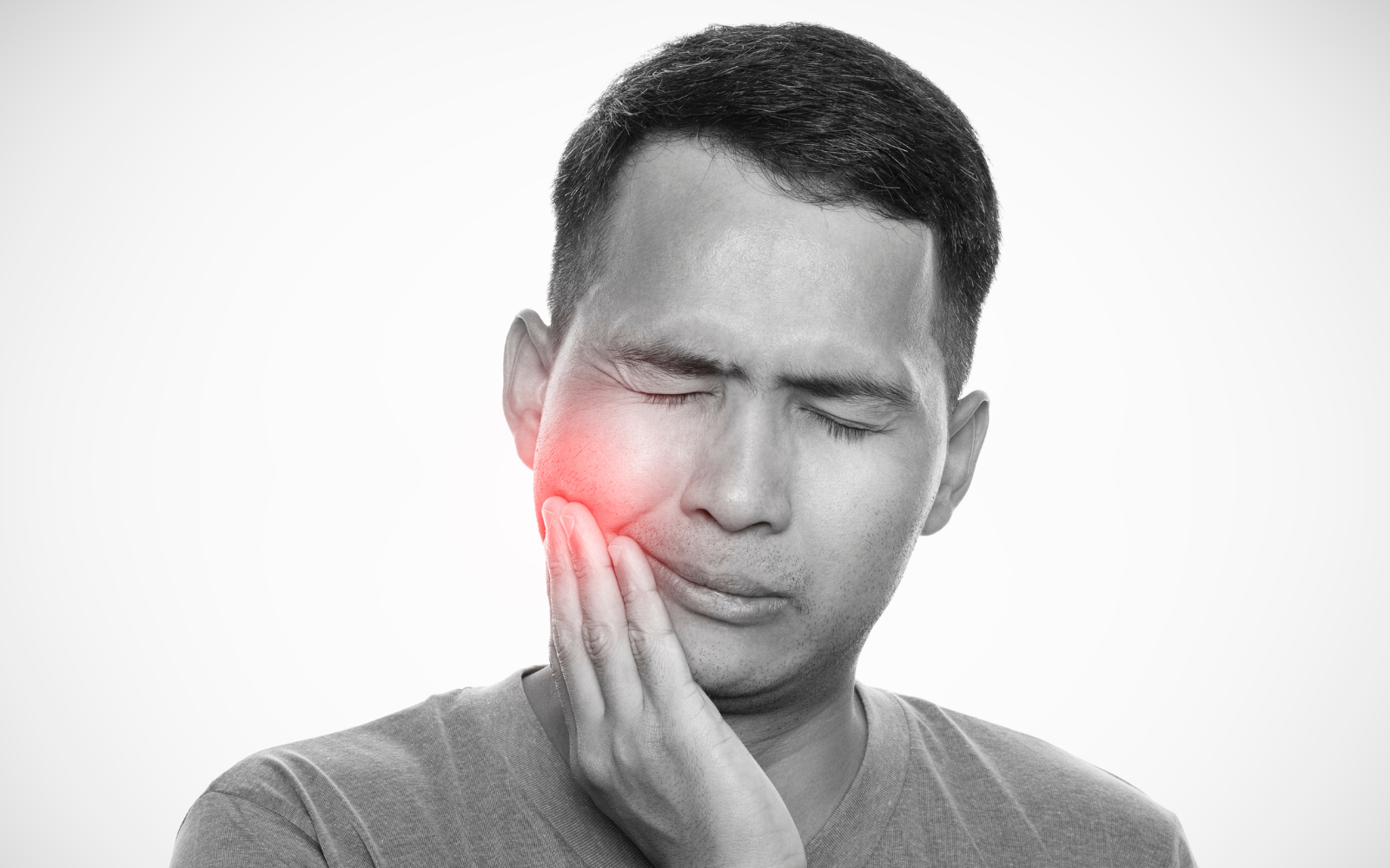
Why does my ear hurt if my ear drum is fine?
Ear pain in TMD almost always has a muscular or articular origin, not an otological one. The masseter, lateral pterygoid, and SCM all have referral patterns that project directly into the ear. The auriculotemporal nerve — which provides sensation to the TMJ — also supplies the external ear. Joint inflammation and trigger point referral can both produce genuine ear pain with a perfectly normal ear drum. If your ENT has cleared you, your jaw muscles and TMJ deserve a proper assessment.
I clench my teeth at night. Is a night guard enough?
A night guard addresses the consequence of bruxism — tooth wear and some joint loading — but does not treat the underlying neuromuscular drivers. Many patients with night guards continue to clench, continue to develop masseter hypertrophy, and continue to have TMJ symptoms because the muscle tension driving the bruxism was never treated. Physical therapy combined with a night guard is significantly more effective than either alone. At Voltex PT, we work alongside your dentist to provide the musculoskeletal component of a comprehensive bruxism management plan.
How many sessions will TMJ treatment take at Voltex PT?
It depends on the severity, chronicity, and complexity of your presentation — but most patients with primary myofascial TMD see meaningful improvement within 4–8 sessions. More complex cases involving disc displacement, cervical spine dysfunction, or long-standing pain sensitization may require 8–16 sessions. We’ll give you an honest assessment and a clear plan at your first visit.
Your Jaw Pain Has a Source. Let’s Find It — and Fix It.
If you’re in Austin and you’re living with jaw pain, facial tension, ear symptoms, TMJ clicking, chronic headaches, or a combination of all of the above — we’re here to help. At Voltex PT, we don’t just treat your jaw. We treat the whole system that’s driving your symptoms — and we do it one-on-one, every session, with doctoral-level clinical expertise.
Stop chasing symptoms. Come find the source.
BOOK YOUR CALL
References
- Peck CC, et al. (2021). Temporomandibular disorders: Current Concepts and Controversies in Diagnosis and Management. PMC. https://pmc.ncbi.nlm.nih.gov/articles/PMC8000442/
- Emedicine/Medscape. TMJ Syndrome: Pathophysiology, Epidemiology. https://emedicine.medscape.com/article/809598-overview
- Physiopedia. Temporomandibular Disorders — DC/TMD Classification. https://www.physio-pedia.com/Temporomandibular_Disorders
- StatPearls. (2025). Anatomy, Head and Neck, Temporomandibular Joint. https://www.ncbi.nlm.nih.gov/books/NBK538486/
- StatPearls. (2023). Anatomy, Head and Neck, Mastication Muscles. https://www.ncbi.nlm.nih.gov/books/NBK541027/
- National Academies of Sciences. (2020). Temporomandibular Disorders: Masticatory System Anatomy and Function. https://www.ncbi.nlm.nih.gov/books/NBK557988/
- Comparative Evaluation of Pain Pattern in Neck Musculature in TMD vs Non-TMD Cases. PMC. https://www.ncbi.nlm.nih.gov/pmc/articles/PMC9642967/
- Altunalan T, et al. (2025). Temporomandibular disorders and neck disability in individuals with cervical disc herniation. European Journal of Oral Sciences. https://pubmed.ncbi.nlm.nih.gov/41157797/
- Effectiveness of Physiotherapy in TMJ Dysfunction and the Relationship with Cervical Spine. PMC. 2022. https://pmc.ncbi.nlm.nih.gov/articles/PMC9687864/
- Neural Basis of Etiopathogenesis and Treatment of Cervicogenic Orofacial Pain. PMC. 2022. https://pmc.ncbi.nlm.nih.gov/articles/PMC9611820/
- Craniocervical Misalignment Masquerading as Facial Pain: SCM and upper trapezius trigger points. CRANIO Journal. 2020. https://www.tandfonline.com/doi/full/10.1080/08869634.2020.1823795
- Correlation between TMD and Cervical Spine Pain and Mobility. PMC. https://www.ncbi.nlm.nih.gov/pmc/articles/PMC4090505/
- Vieira LS, et al. (2023). The Efficacy of Manual Therapy Approaches on Pain, Mouth Opening and Disability in TMD: Systematic Review of 20 RCTs. Life. 13(2):292. https://doi.org/10.3390/life13020292
- Upper Cervical Mobilization/Manipulation on TMJ Pain, Maximal Mouth Opening, and Pressure Pain Thresholds: Systematic Review and Meta-Analysis. PMC. 2023. https://pmc.ncbi.nlm.nih.gov/articles/PMC10036235/
- Khayamzadeh M, Razmara F, Tavassoli A. (2025). Dry Needling in Treatment of TMJ Disorders: A Systematic Review. Clin Exp Dent Res. PMID:40917038. https://pubmed.ncbi.nlm.nih.gov/40917038/